Tendon Diagram Labeled - Sports Injuries To The Hand Wrist Elbow Ski Tennis Golf - The subacromial bursa lies between the rotator cuff and shoulder blade and protects the tendons in this area.
Tendon Diagram Labeled - Sports Injuries To The Hand Wrist Elbow Ski Tennis Golf - The subacromial bursa lies between the rotator cuff and shoulder blade and protects the tendons in this area.. If you're looking for a speedy way to learn muscle anatomy, look no further than our anatomy crash courses. The smaller bone that runs alongside the tibia (fibula) and the. Posted on february 14, 2015 by admin. They may occur suddenly during activity, or gradually over time. Allows the action of raising the foot.
In the back and elsewhere in the body, tendons attach muscles to bones. The smaller bone that runs alongside the tibia (fibula) and the. Tendons] connective tissue structures identifiable in gross anatomy: As these muscles contract and relax, they move skeletal bones to create movement of the body. See muscle contraction diagram stock video clips.
Https Www Uc Edu Content Dam Uc Ce Images Olli Page 20content Muscular 20system 20s Pdf from In human anatomy, the peroneus longus (also known as fibularis longus) is a superficial muscle in the lateral compartment of the leg, and acts to evert and plantarflex the ankle. Arm muscle diagram labeled simple : However, the long head of the biceps brachii is one of the more common tendons to rupture. Further, some ligaments prevent movement in. A muscle's origin is where a tendon attaches it to the *less* movable bone. Human anatomy diagrams show internal organs, cells, systems, conditions, symptoms and sickness information and/or tips for healthy living. Black and white print showing the musculoskeletal system of a human hand, including the bones, muscles, cartilage, tendons, ligaments, and joints,. Thank you and have a good time!
This diagram depicts muscle structure diagram.
Skeletal muscle diagram muscle fascia heart development types muscles fascia human body muscle and fascia heart cell fascia skeletal muscle cell anatomy muscular contraction. Browse 318 hand anatomy tendons stock photos and images available, or start a new search to explore more stock photos and images. Achilles tendon the achilles tendon is a band of tissue that connects a muscle to a bone. All these diagrams are free and printable, just click on the picture to enlarge and save it. The patient would not notice much weakness in the upper limb due to the. The tendons have 2 functions: Three of them are located in the anterior compartment — the biceps brachii, brachialis, and coracobrachialis, while the forth is located in the posterior compartment — the triceps brachii). In the diagram of the humerus this structure receives the head of the radius when the forearm is flexed. A muscle's origin is where a tendon attaches it to the *less* movable bone. See muscle contraction diagram stock video clips. A complete rupture of any tendon in the body is rare. When autocomplete results are available use up and down arrows to review and enter to select. The smaller bone that runs alongside the tibia (fibula) and the.
Muscle diagram blank each of these muscles is a discrete organ constructed of skeletal muscle tissue, blood vessels, tendons, and nerves. Human anatomy diagrams show internal organs, cells, systems, conditions, symptoms and sickness information and/or tips for healthy living. Rupture of the biceps tendon. The muscle belly then crosses the entire upper arm and separates into two tendons. As these muscles contract and relax, they move skeletal bones to create movement of the body.
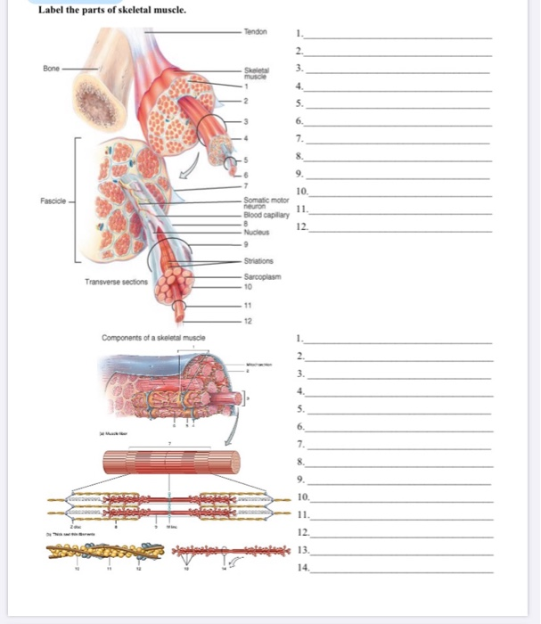
Label The Parts Of Skeletal Muscle Tendon 1 2 Bone Chegg Com from media.cheggcdn.com In the diagram of the humerus this structure receives the head of the radius when the forearm is flexed. Muscular system anatomy and physiology nurseslabs : The muscle belly then crosses the entire upper arm and separates into two tendons. Browse 318 hand anatomy tendons stock photos and images available, or start a new search to explore more stock photos and images. Three of them are located in the anterior compartment — the biceps brachii, brachialis, and coracobrachialis, while the forth is located in the posterior compartment — the triceps brachii). If you're looking for a speedy way to learn muscle anatomy, look no further than our anatomy crash courses. Muscle diagram blank each of these muscles is a discrete organ constructed of skeletal muscle tissue, blood vessels, tendons, and nerves. Shoulder tendons chart ~ labeled anatomy chart of shoulder ligaments on white background stocktrek images.
{label gallery} get some ideas to make labels for bottles, jars, packages, products, boxes or classroom activities for free.
The smaller bone that runs alongside the tibia (fibula) and the. The knee is one of the largest and most complex joints in the body. While multiple groups exists, overall, there are 3 different types of muscle tissues: The subacromial bursa lies between the rotator cuff and shoulder blade and protects the tendons in this area. To understand one of the most complex joints of our body i.e. Muscular system anatomy and physiology nurseslabs : Attaches the calf muscles to the calcaneus, most important muscles for running, jumping, walking etc. They may occur suddenly during activity, or gradually over time. Make writing personal training programs easy with these custom designed exercise templates, and keep your clients focused and progressing. Skeletal muscle diagram muscle fascia heart development types muscles fascia human body muscle and fascia heart cell fascia skeletal muscle cell anatomy muscular contraction. Allows the foot to be turned inward and also supports the arch of the foot. We provide you with the unlabeled version for evaluation. Achilles tendon the achilles tendon is a band of tissue that connects a muscle to a bone.
While multiple groups exists, overall, there are 3 different types of muscle tissues: {label gallery} get some ideas to make labels for bottles, jars, packages, products, boxes or classroom activities for free. Study the overall and complete anatomy of the leg muscle using these diagrams. It runs down the back of the lower leg and connects the calf muscle to the. Diagram of tendons in forearm 👉 we are pleased to provide you with the picture named right arm muscle and tendon anatomywe hope this picture right arm muscle and tendon anatomy can help you study and research.
Muscle Diagram Muscular System Human Muscular System Muscular System Labeled from i.pinimg.com One or more ligaments provide stability to a joint during rest and movement. A tendon is a structure that connects muscle to bone, and the biceps are connected by tendons at both the elbow and shoulder joints. The muscle belly then crosses the entire upper arm and separates into two tendons. 19 photos of the knee tendon anatomy diagram and name chart. When autocomplete results are available use up and down arrows to review and enter to select. {label gallery} get some ideas to make labels for bottles, jars, packages, products, boxes or classroom activities for free. This will help you to understand the mechanism as well as the working. Muscle diagrams are a great way to get an overview of all of the muscles within a body region.
As these muscles contract and relax, they move skeletal bones to create movement of the body.
Also allows the action of raising up onto toes. The subacromial bursa lies between the rotator cuff and shoulder blade and protects the tendons in this area. Muscular system anatomy and physiology nurseslabs : A muscle's origin is where a tendon attaches it to the *less* movable bone. Muscle charts of the human body. Huesos del miembro superior arm anatomy arm bones human anatomy The hand incorporates countless muscles, bones, tendons and ligaments into simple motion and this chart covers them all. They may occur suddenly during activity, or gradually over time. In the diagram of the humerus this structure receives the head of the radius when the forearm is flexed. The knee is one of the largest and most complex joints in the body. If you're looking for a speedy way to learn muscle anatomy, look no further than our anatomy crash courses. While multiple groups exists, overall, there are 3 different types of muscle tissues: However, the long head of the biceps brachii is one of the more common tendons to rupture.


0 Komentar